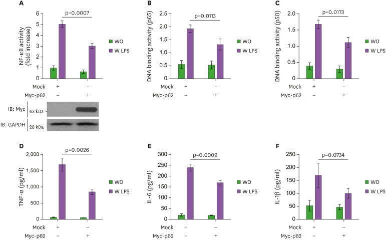
Figure 1. p62 overexpression inhibits the activation of NF-κB induced by TLR4. (A) THP-1 cells were transfected with vector control (Mock) or Myc-p62 vector together with pBIIx-luc and Renilla luciferase vector, untreated or treated with LPS (200 ng/ml) for 6 h, then subjected to the luciferase activity assay. The results are expressed as fold-induction in luciferase activity relative to untreated cells. All error bars represent the mean ± SD of triplicate samples. Western blot analysis was used to measure the expression of transiently transfected Myc-p62 (lower panel). The expression of GAPDH was used as a loading control. (B and C) THP-1 cells were transfected with mock or Myc-p62 vector, untreated or treated with LPS (200 ng/ml) for 6 h, then analyzed for p65-DNA (B) or p50-DNA (C) binding activity. All error bars represent the mean ± SD of triplicate samples. (D-F) THP-1 cells were transfected with mock or Myc-p62 vector, untreated or treated with LPS (200 ng/ml) for 9 h, then subjected to ELISA to determine the levels TNF-α (D), IL-6 (E), and IL-1β (F) produced. All error bars represent the mean ± SD from triplicate samples.
WO, untreated; W LPS, treated with LPS.

