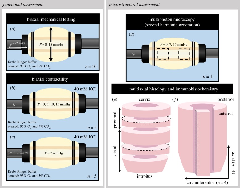
Figure 1.
Schematic for functional and microstructural assessment of murine vaginal tissue. Tissues were subjected to extension–inflation protocols (a) for biaxial mechanical testing. Biaxial contractile testing was executed under various constant pressures (b) and fixed lengths (c). Multiphoton microscopy with second harmonic generation was performed at the averaged physiological length along the anterior wall at the proximal and distal regions under three constant pressures (d). Histological and immunohistochemical analysis was performed on circumferential sections taken from the proximal and distal regions of the vaginal wall (e); axial sections were taken along the length of the anterior wall (f). The dashed lines represent the region of interest. (Online version in colour.)

