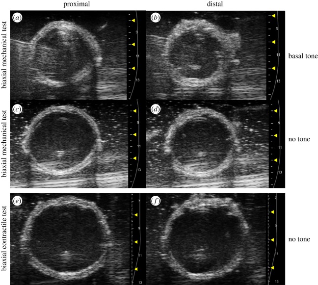
Figure 2.
Representative ultrasound B-mode images acquired at the proximal (a,c,e) and distal (b,d,f) regions of the vaginal wall. During biaxial mechanical testing (a–d), images were acquired at the unloaded configuration with (a,b) and without (c,d) basal tone. For biaxial contractile testing (e,f), images were acquired at the unloaded configuration without muscle tone. (Online version in colour.)

