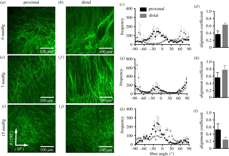
Figure 6.
Multiphoton microscopy with SHG of the collagen fibre architecture at the proximal (a,e,i) and distal (b,f,j) regions of the anterior vaginal wall under 0 (a,b), 7 (e,f) and 15 (i,j) mmHg at the average physiological length. The frequency of the number of individual fibre segments with its respective orientation is reported with 0° denoting the axial axis and ±90° denoting the circumferential (c,g,k) at increments of 5°. At 0 and 7 mmHg, collagen fibres are oriented towards the circumferential axis at the distal region. At the proximal region, fibres are oriented towards the circumferential and diagonal axes (c,g). Under 15 mmHg, at the proximal and distal regions, the fibres are oriented towards the axial direction (k). The alignment coefficient describes the collagen fibre orientation towards a preferred direction, with 1 being highly aligned (d,h,l). The alignment coefficient was larger for the distal region than for the proximal at 0 and 7 mmHg. Data were averaged over several images and are reported as mean ± s.e.m. Imaging depth was 7–10 µm from the adventitial layer or outer wall. (Online version in colour.)

