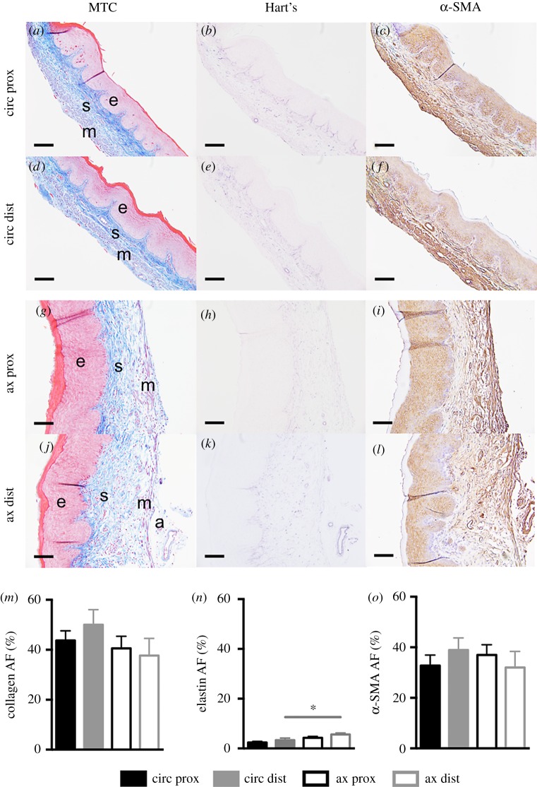
Figure 7.
Representative histological images were acquired at 20× magnification. Proximal (prox; a–c, g–i) and distal (dist; d–f, j–l), circumferential (circ; a–f) and axial (ax; g–l) sections stained with Masson's trichrome (MTC; a,d,g,j), Hart's elastin (b,e,h,k) and α-SMA (c,f,i,l). Layers of the vaginal wall are denoted: epithelium (e), subepithelium (s), muscularis (m) and adventitia (a). Area fraction (AF) for collagen (m), elastin (n) and α-SMA (o) are reported for the proximal (black) and distal (grey) regions along the circumferential (close) (filled) and axial (open) sections. At the distal region, the elastin area fraction was greater for the axial section than for the circumferential. Data are reported as mean ± s.e.m. Statistical significance is denoted by *p < 0.05/2. Scale, 100 µm. (Online version in colour.)

