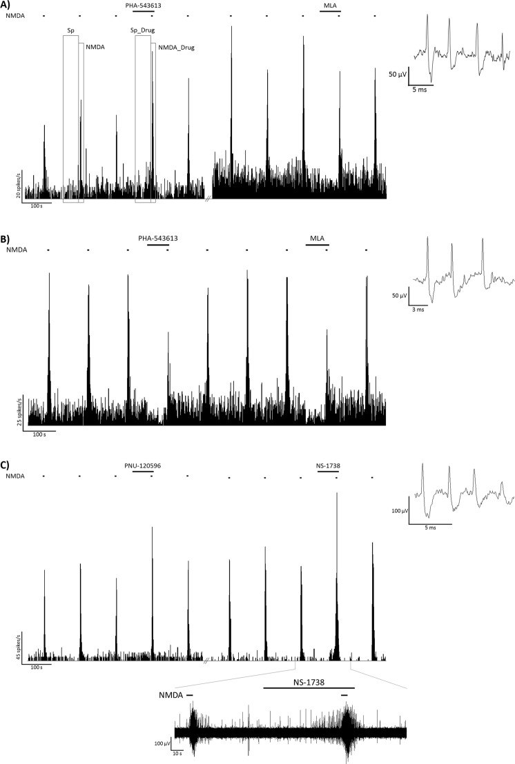
Figure 1.
Firing rate histograms from representative recordings of the local effects of PHA-543613 and MLA (A,B), and PNU-120596 and NS-1738 (C) on the spontaneous and NMDA-evoked firing activity of CA1 hippocampal pyramidal neurons. Furthermore, panel (A) shows the time windows for calculating spontaneous and NMDA-evoked firing rates before (Sp, NMDA, respectively), and during (Sp_Drug, NMDA_Drug) microiontophoretic application of a given test compound. Insets to the right show a typical extracellular action potential recorded in the given experimental session. Bottom inset on panel (C) shows the raw electrophysiological recording in the time window marked on the horizontal axis of the corresponding histogram.

