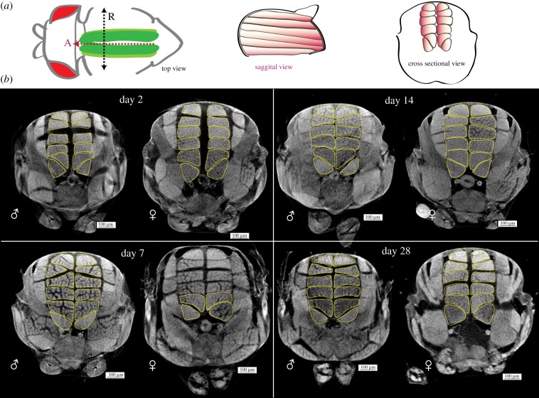
Figure 1.
Gross changes in male and female adult Drosophila longitudinal muscles over time. (a) Schematic of dorsal longitudinal muscle (DLM) positioning in the adult thorax. In top view, DLMs (green) run along the A–P axis (red dotted line, arrowhead indicates anterior ‘A’) inside the thorax, under the cuticle. The black dotted line describes the left–right axis, running between the wing hinges. ‘R’ denotes the animal's right-hand side. The sagittal view shows six muscle fibres (orange) run anterior to posterior in one hemithorax. In cross-sectional view, six DLMs are arranged in the thorax, on either side of the midline. (b) Representative cross sections of whole thorax microCT scans of male (♂) and female (♀) flies at days 2, 7, 14 and 28 post-eclosion. DLMs outlined with yellow dotted lines. Scale bars, 100 µm; n = 14–21 per sex per time point.

