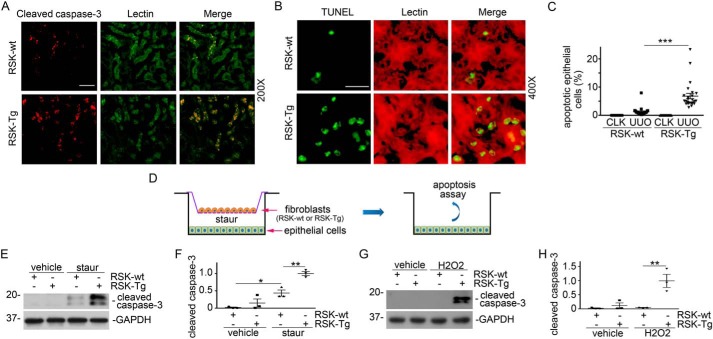
Figure 3.
p90RSK induces fibroblast-mediated tubular epithelial apoptosis in vivo and in vitro. A, obstructed kidney sections from FSP-1–specific p90RSK transgenic mice (RSK-Tg) and their littermates (RSK-wt) were subjected to immunofluorescence staining of cleaved caspase-3 (red) and lectin (green, proximal tubular epithelial marker), 200×. Scale bar, 50 μm. B, immune staining of TUNEL (green) and lectin (red), 400×. Scale bar, 20 μm. C, quantitation of apoptotic epithelial cells. ***, p < 0.001; n = 5 microscopic fields per mouse × 5 mice/group. D, illustration of primary fibroblast (RSK-Tg and RSK-wt) and epithelial cell (HKC-8) coculture. After serum starvation overnight in the coculture, 50 nm staurosporine or 150 μm H2O2 was added to the lower chamber for additional 4 or 16 h, respectively. Then, HKC-8 cells were harvested and subjected to Western blotting for cleaved caspase-3 and GAPDH. E, Western blotting of cleaved caspase-3 after staurosporine treatment. F, quantitation of cleaved caspase-3 abundance. *, p < 0.05; **, p < 0.01; n = 3 experiments. G, Western blotting of cleaved caspase-3 after H2O2 treatment. H, quantitation of cleaved caspase-3 abundance. **, p < 0.01; n = 3 experiments. CLK, control kidney; staur, staurosporine. Error bars, S.E.

