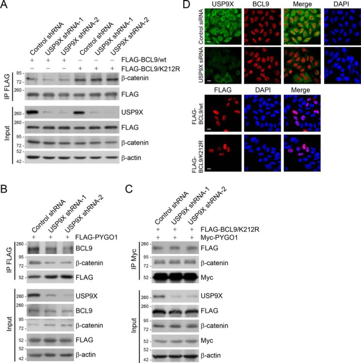
Figure 3.
USP9X-promoted BCL9 deubiquitination controls the formation of β-catenin/BCL9/PYGO1 complex. A, HeLa cells stably expressing different sets of USP9X shRNAs and FLAG–BCL9/WT or FLAG–BCL9/K212R were collected. The cellular extracts were prepared and analyzed with IP followed by IB with antibodies against the indicated proteins. B, HeLa cells stably expressing different sets of USP9X shRNAs were transfected with FLAG–PYGO1. The cellular extracts were prepared and analyzed with IP followed by IB with antibodies against the indicated proteins. C, HeLa cells stably expressing different sets of USP9X shRNAs were transfected with Myc–PYGO1 and FLAG–BCL9/K212R. The cellular extracts were prepared and analyzed with IP followed by IB with antibodies against the indicated proteins. D, immunostaining and confocal microscopy analysis of USP9X and BCL9 subcellular localization in USP9X-depleted HeLa cells (upper panel). Immunostaining and confocal microscopy analysis of the subcellular localization of FLAG–BCL9/WT and FLAG–BCL9/K212R in HeLa cells (lower panel) are shown. Scale bar, 10 μm.

