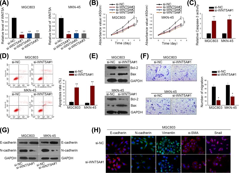
Figure 3. WNT5A functioned as oncogenes in regulating GC biological process.
(A) WNT5A expression was silenced by si-WNT5A#1/2/3 in both MGC803 and MKN-45 cells. (B) CCK-8 assay was used to measure GC cell proliferation after silencing WNT5A expression. (C) Caspase-3 activity was detected for GC cell apoptosis. (D) Apoptotic cells in MGC803 and MKN-45 cells were determined by flow cytometry. (E) Western blot analysis for apoptosis-related proteins (Bcl-2 and Bax). (F) GC cell migratory ability was evaluated by Transwell migration assay. (G) Western blotting for EMT-associated proteins (E-cadherin and N-cadherin). (H) Immunofluorescence staining examined the different expression levels of EMT biomarkers MGC803 cells transfected with si-NC or si-WNT5A#1. **P < 0.01 and *P < 0.05 vs control group.

