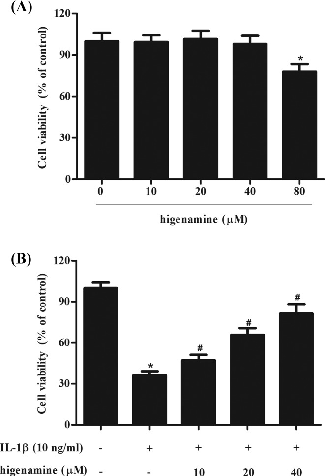
Figure 1. Effect of higenamine on cell viability in NPCs exposed to IL-1β.
(A) NPCs were incubated with different concentrations of higenamine (0, 10, 20, 40, and 80 μM) for 24 h. MTT assay was performed to evaluate cell viability. (B) NPCs were pretreated with 10, 20, 40 μM of higenamine for 2 h, followed by the induction of IL-1β (10 ng/ml) for 24 h. Cell viability was detected using MTT assay. *P < 0.05 vs. control NPCs. #P < 0.05 vs. IL-1β-stimulated NPCs.

