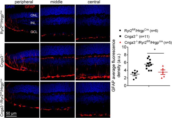
Figure 7.

Deletion of Ryr2 reduced activation of Müller glial cells in Cnga3-/- mice. GFAP immunofluorescence labeling was performed on the retinal cross sections prepared from Cnga3-/-/Ryr2flox/flox/Hrgp-cre, Cnga3-/-, and Ryr2flox/flox/Hrgp-cre mice at P30. Shown are representative confocal images of immunofluorescence labeling of GFAP on the peripheral, middle, and central regions of the retinal sections and corresponding quantification of immunofluorescence intensity. RGC, Retinal ganglion cell. Data are presented as mean ± SEM. *p < 0.05.
