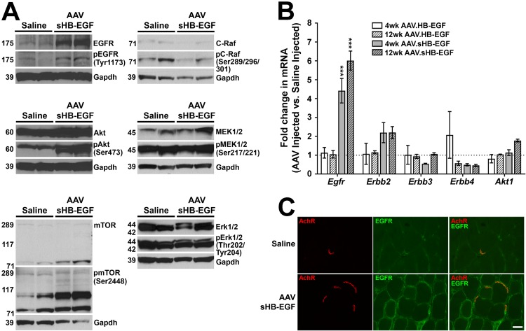
FIG 6.
sHB-EGF stimulates an EGFR-Akt signaling pathway in skeletal muscle tissue. (A) Western blots of protein lysates from muscles treated for 4 weeks with r(sc)AAV9.CMV.sHB-EGF and saline-treated contralateral control muscles probed using antibodies to GAPDH protein (a protein loading and transfer control), antibodies to EGFR, Akt, mTOR, C-Raf, MEK1/2, or Erk1/2 protein, or antibodies specific to phosphorylated EGFR (at Tyr1173), Akt (at Ser473), mTOR (at Ser2448), C-Raf (at Ser289/296/301), MEK1/2 (at Ser217/221), or Erk1/2 (at Thr202/Tyr204). (B) qRT-PCR measures of Egfr, Erbb2, Erbb3, Erbb4, and Akt1 gene expression in muscles from mice treated for 4 or 12 weeks with r(sc)AAV9.CMV.sHB-EGF or r(sc)AAV9.CMV.HB-EGF relative to levels for saline-injected control muscles (set to 1). Errors are SEM for n = 4 measures per condition. *, P < 0.05; **, P < 0.01; ***, P < 0.001. (C) Wild-type saline-injected or r(sc)AAV9.CMV.sHB-EGF-injected muscles were stained with rhodamine-conjugated α-bugarotoxin to identify nicotinic acetylcholine receptors (AChRs) (red) at the neuromuscular junction and with an antibody to EGFR (green). Merged images on the right show overlapping staining in orange and/or yellow. Scale bar is 25 μm.

