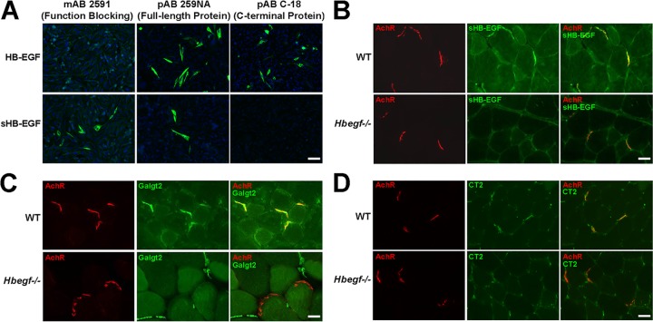
FIG 7.
Enrichment of sHB-EGF at the neuromuscular synapse. (A) CHO cells were transfected with cDNAs expressing either soluble HB-EGF (sHB-EGF) or full-length transmembrane HB-EGF. Cells were then immunostained with MAb 2591 (an sHB-EGF-specific function blocking antibody), PAb 259 NA (recognizes full-length HB-EGF protein as well as sHB-EGF), or PAb C18 (recognizes the C-terminal intracellular domain of full-length HB-EGF). Nuclear (DAPI) staining is shown in blue. Scale bar is 50 μm. WT and HB-EGF-deficient (Hbegf−/−) muscle sections were stained with the sHB-EGF-specific antibody MAb 2591 (B), a Galgt2 antibody (C), or an antibody to CT glycan (CT2) (D) (all shown in green). Tissue sections were costained with rhodamine-conjugated α-bungarotoxin (red) to label acetylcholine receptors (AChRs) at the neuromuscular junction. Merged images at the right show staining overlap in yellow or orange. Scale bar is 25 μm.

