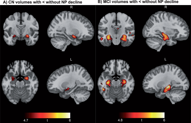
Fig.3.
Neuropsychological decline linked to baseline hippocampal and medial temporal lobe volume. Results of voxel-based morphometry analysis after FWE correction are displayed as t-maps overlaid onto a template T1 image for anatomical reference. Findings from two-sample t-tests show a consistent pattern of smaller hippocampal volume in CN participants with NP decline versus CN without NP decline (A) and smaller hippocampal and medial temporal volume in MCI patients with NP decline versus MCI without NP decline (B).

