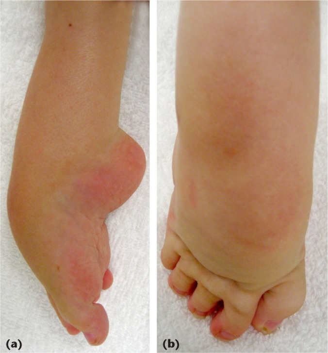
Fig. 2.

Equinocavus clubfoot variant: (a) lateral view of the left foot depicting the severe hindfoot equinus and deep transverse cavus. The toes are mildly flexed in this uncorrected foot; (b) dorsal view of the same foot, showing relatively mild forefoot adductus
