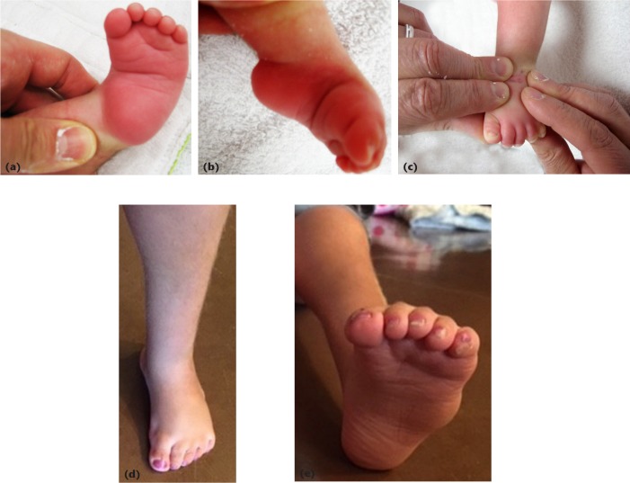
Fig. 5.
Equinocavus clubfoot variant in a two-month-old girl: (a) plantar view of left foot, demonstrating medial to lateral plantar crease; (b) medial view showing severe ankle and midfoot equinus, with plantarflexed great toe; (c) the ‘four finger’ technique, the index and long fingers of both hands are positioned over the midfoot to act as a fulcrum, while the thumbs apply dorsiflexion pressure under the heads of the metatarsals. The intact Achilles tendon acts as a counter force, therefore, an Achilles tenotomy should be delayed until full correction of the midfoot cavus; (d) the same foot dorsal view, weight-bearing at seven years’ follow-up; (e) plantar view. The only other treatment she required was percutaneous toe flexor tenotomies at three years of age.

