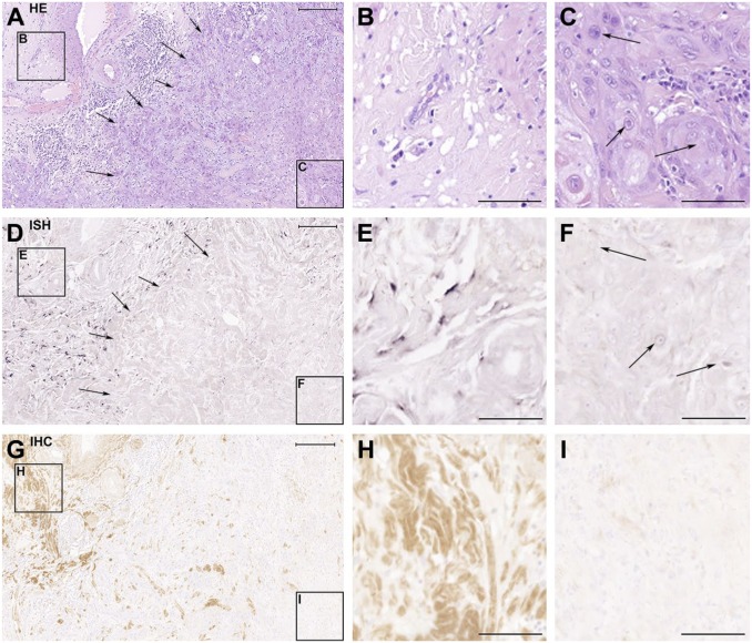
Figure 1.
Human vulva carcinoma cells are negative for decorin expression in vivo. The panel consists of serial sections of a representative human vulva carcinoma tissue sample analyzed with hematoxylin and eosin staining (A-C), ISH for decorin (D-F), and IHC for decorin (G-I). The frames on the left side of the panels A, D, and G mark non-malignant tissue areas, shown magnified in panels B, E, and H, respectively. The frames on the right side of the panels A, D, and G mark carcinoma areas, and they are shown magnified in panels C, F, and I, respectively. Arrows in A and D mark the border between malignant and non-malignant tissue areas. Arrows in C and F point to squamous carcinoma cells. Positive ISH signal for decorin can be seen in purple (D and E). Positive immunostaining for decorin can be seen in brown in G and H. Note that there is a total absence of decorin mRNA expression and immunoreactivity in panels F and I that represent tissue areas populated by malignant cells. Scale bar in A, D, and G 500 µm and in B, C, E, F, H, and I 200µm. Abbreviations: HE, hematoxylin and eosin; ISH, in situ hybridization; IHC, immunohistochemistry.

