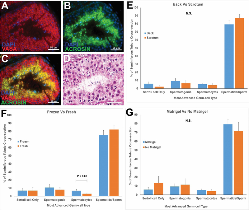
Figure 5: Histological evaluation of spermatogenic development in grafts.

Immunofluorescence staining of recovered grafted tissue for VASA+ germ cells (red, A) and ACROSIN+ post-meiotic cells (green, B). DAPI counterstain marks all cell nuclei (blue). The merged VASA/ACROSIN/DAPI co-stain is shown in (C). Please see Figure S4 for additional markers of undifferentiated spermatogonia (UTF1), spermatocytes (BOULE) and spermatids (CREM). Hematoxylin and Eosin staining of post-graft tissues (D). Please see H and E staining for grafts from each individual animal in Figure S5. Quantification of most advanced germ cell type in graft seminiferous tubules (E-G). Bars represent mean ± standard error of the mean. N.S. = not significant; P<0.05 was considered statistically significant.
