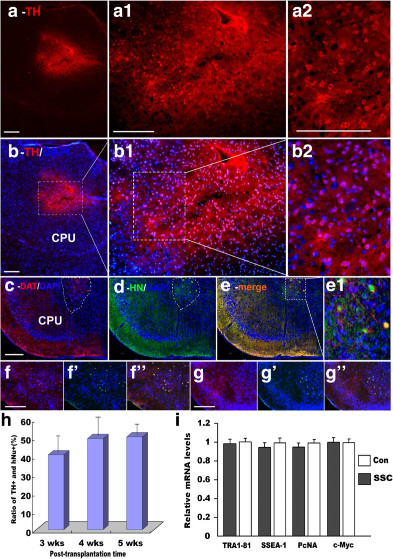
Fig. 7.

Transplantation of hSSCs into the striatum of mice and examination of tumorigenesis (a) TH antibody marks hSSC-derived cells integrated into the mouse brain. a1 shows a magnification of a. a2 shows a magnification of (a1). b–b2 DAPI counterstaining of a–a2). c, d Immunofluorescence of analyses using another specific antibody targeted against DA neuron-specific DAT (c) and human Nuclei (hNuc) antibody (d). e Double-staining of DAT and hNuc. e1 shows a magnification of e. f–f”, g–g” double-immunostaining of nestin (f, g) with cyclinD1 (f’, g’) at 1 and 2 weeks posttransplantation, respectively. h Quantification of the differentiation potential of DA neurons at 3, 4, and 5 weeks posttransplantation. Approximately 40–50% of the hSSC-derived cells stain TH-positive when transplanted into the striatum of mice for 3, 4 and 5 weeks. i Quantitative RT-PCR analysis of key genes as indicated. Scale bars = 50 μm
