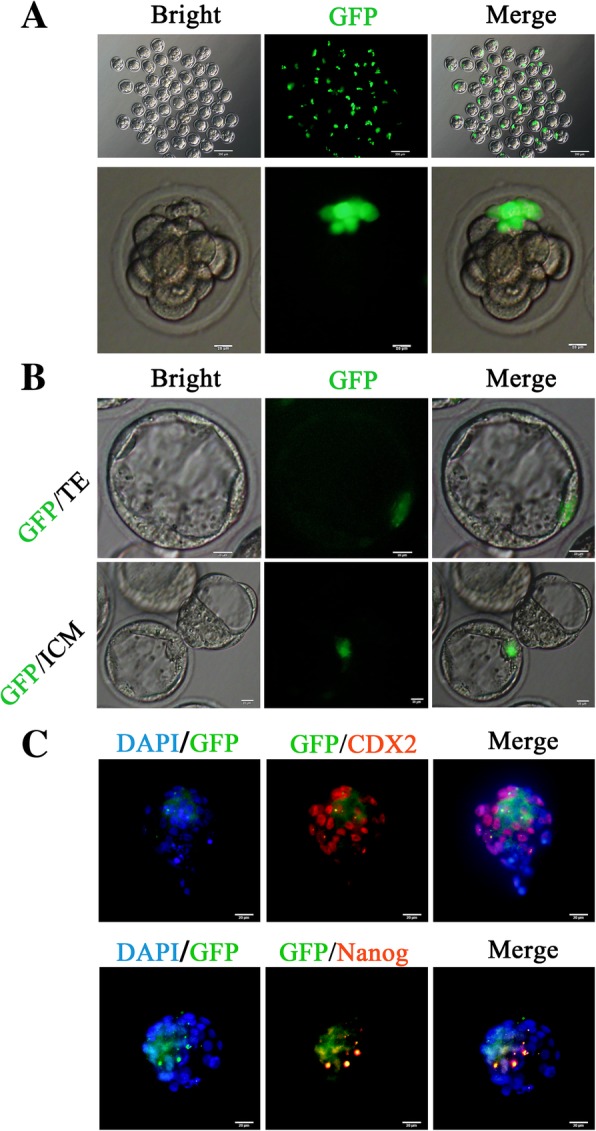
Fig. 4.

PC-iPS contributes to mouse late blastocyst TE and ICM in vitro. a Injection GFP labeled PC-iPS cells to 4- to 8-cell embryos, scale bar 200 μm, 20 μm. b GFP-labeled PC-iPS cells contribute to the TE and ICM, scale bar 20 μm. c Immunocytochemistry analysis of CDX2 and NANOG in chimeric mouse late blastocyst, scale bar 20 μm
