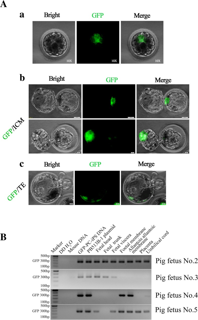
Fig. 6.

GFP-labeled PC-iPS cells contribute to chimeric formation in pig embryos in vitro and in vivo. (A, a) Representative fluorescence images of pig embryo injected with GFP-labeled PC-iPS cells, scale bar 10 μm. (A, b) GFP-labeled PC-iPS contribute to the pig ICM, scale bar 50 μm. (A, c) GFP-labeled PC-iPS cells contribute to the pig TE, scale bar 50 μm. (B) Nested PCR analysis of GFP using post-implantation pig fetal embryonic and extraembryonic tissues (the GFP-labeled PC-iPS cells’ DNA and PB513B-1 plasmid were used as GFP sequence positive controls; double distilled water (ddH2O) and mouse DNA were used as negative controls)
