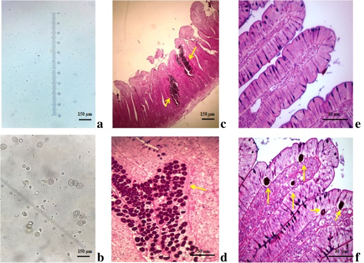
Fig. 1.
Oocysts detection in the feces of unchallenged, UC a and Eimeria-challenged, EC b broiler chickens 144 h post-inoculation (PI). c, d Histological images of duodenal villi and e, f jejunal villi in broiler chickens 144 h PI, 40× magnification. Note the presence of Eimeria structures (yellow arrows) in the duodenal and jejunal mucosa of EC broilers. e Intact jejunal villi in UC broilers. Hematoxylin-eosin staining. a, b, c Scale bars represent 150 μm. d, e, f Scale bars represent 30 μm

