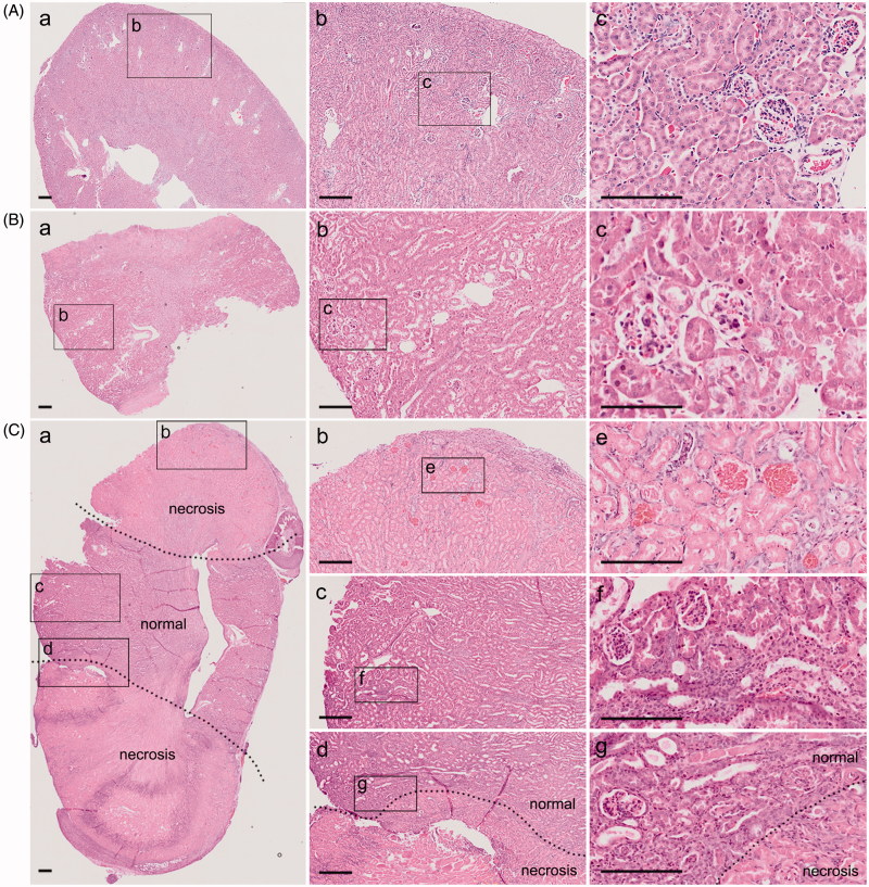
Figure 2.
Unilateral kidney ligation simulates conventional 5/6 nephrectomy. (A) H&E staining of kidney in sham group after surgery 1 week ((Aa) is a × 40 image, (Ab) is a magnified image from (Aa) with ×100 magnification, and (Ac) is a magnified image from (Ab) with ×200 magnification). (B) H&E staining of kidney in c-PNx group after surgery 1 week ((Ba) is a × 40 image, (Bb) is a magnified image from (Ba) with ×100 magnification, and (Bc) is a magnified image from (Bb) with ×200 magnification). (C) H&E staining of kidney in l-PNx group after surgery 1 week ((Ca) is a × 40 image, and (Cb) is necrotic portion, Cc is normal portion, and Cd is junction portion with ×100 magnification, (Ce,Cf,Cg) is magnified image from (Cb,Cc,Cd) with ×200 magnification).

