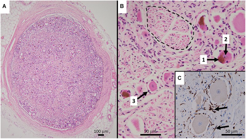
FIGURE 1.

(A) Representative HE stained micrograph section of a thoracic human DRG (medical dissection course, ethics approval obtained from The Southern Adelaide Clinical Human Research Ethics Committee, OFR no.: 55.17) with a thick protective layer of connective tissue demonstrating the predominant localization of cell bodies in the periphery of the ovoid DRG cross-section. (B) Cell bodies of sensory neurons containing lipofuszin (1) and a nucleus with a prominent nucleolus (2), surrounded by satellite cells (3). Bundles of nerve fibers (dashed line) are predominantly present in the center of the ganglion. The HE staining method results in shrinkage of the cell bodies which disconnects them from the layer of satellite cells. (C) Immunohistochemistry micrograph for CD163 with counterstaining for hematoxylin shows the presence and distribution of macrophages (arrows) in DRG.
