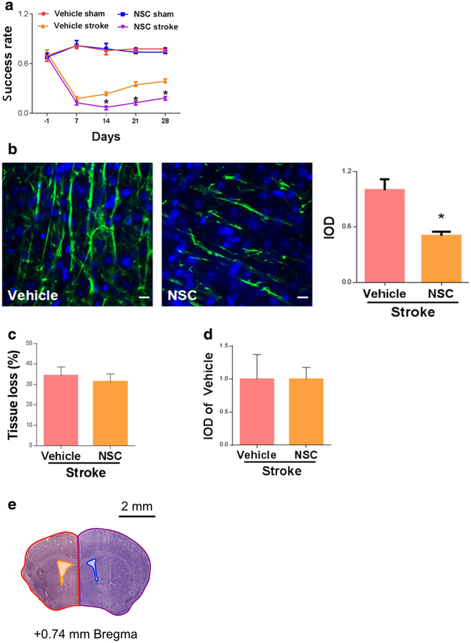
Fig. 1.
Delayed inhibition of Rac1 led to poorer recovery and reduced axonal density after stroke. a Functional recovery assessed by pellet reaching test from day 14 to day 28 after stroke. N = 7–9, *p < 0.05. b Axonal density (neurofilament staining)in the peri-infarct zone was assessed on 28 days after stroke. Scale bars: 10 μm. N =4,*p < 0.05. c NSC treatment did not affect tissue loss 28 days after stroke. N =7–9, p > 0.05. d NSC treatment did not reduce BrdU-positive cells at 28 days after stroke. N =4, p > 0.05. e An illustration of how quantification of brain tissue loss was performed after stroke using CV staining. % brain tissue lost = 100% × [(contralateral hemisphere area - contralateral ventricular area) - (ipsilateral hemisphere area - ipsilateral ventricular area)]/(contralateral hemisphere area - contralateral ventricular area). Red area: ipsilateral hemisphere area; yellow area: ipsilateral ventricular area; purple area: contralateral hemisphere area; blue area: contralateral ventricular area. A specific Rac1 inhibitor, NSC23766 (NSC), or vehicle (saline) was administrated to WT mice starting 7 days after MCAO and injected once daily for 7 days (4.0 mg/kg/day). Data were as mean ± SEM

