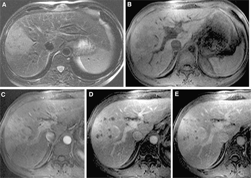
Fig. 2.
50-year-old male with cHCC-CC. There was a consensus achieved among readers regarding moderate high signal intensity on T2-weighted imaging (A) and arterial phase hyperenhancement (C). However, there was no consensus regarding liver surface retraction, presence of progressive enhancement (D, E), tumor in vein, or LI-RADS score (2 scores of LR-M, 1 score as LR-4, and 1 as LR-5 V). The patient underwent partial hepatectomy and the final diagnosis was cHCC-ICC.

