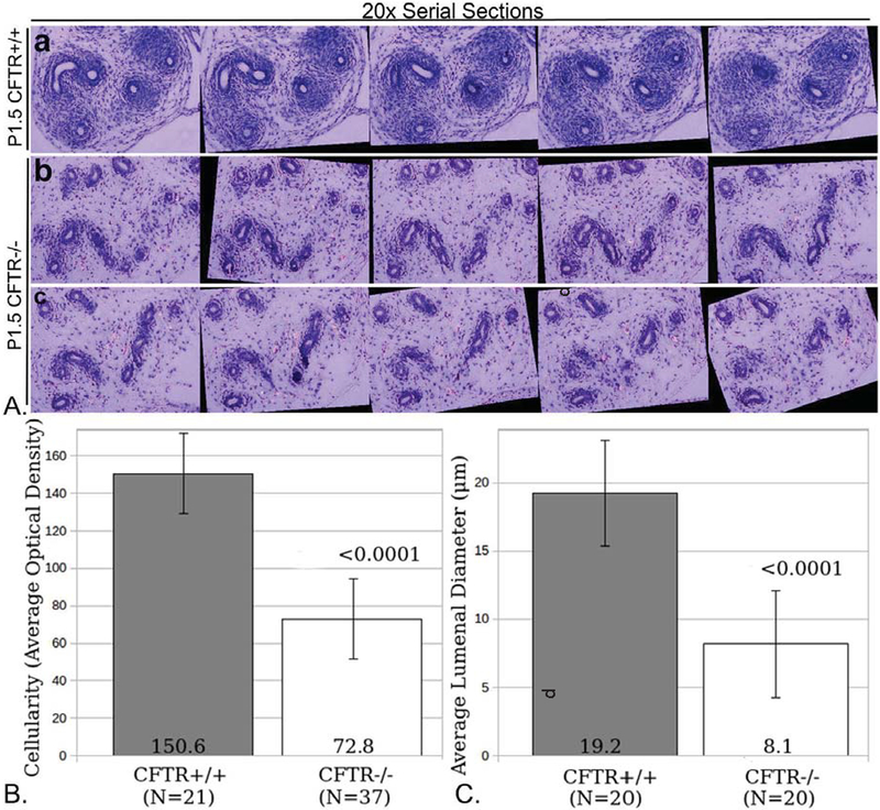
Figure 5: Reduced cellularity and luminal diameter of caput epididymis in two-day-old CF rat.
(Panel A) Serial sections from caput epididymis of CFTR+/+ (a) or CFTR−/− (b,c) animals were stacked and aligned using ImageJ (Methods) to visualize abnormal development of CFTR−/− epididymis. Note lack of smooth muscle proliferation surrounding CFTR−/− caput epididymal ducts (see also Figure 7). (Panel B) ImageJ analysis of optical density (O.D.) showing reduced cellularity external to epithelial duct. Cellular densities immediately surrounding 21 wild type and 37 CFTR null ducts were measured. (Panel C) Reduced lumenal diameter was observed in the caput epididymis of CFTR−/− rats at birth. To obtain duct diameter, 20 wild type and 20 CF ducts were evaluated (from 3 animals per group). Error bars= SEM.

