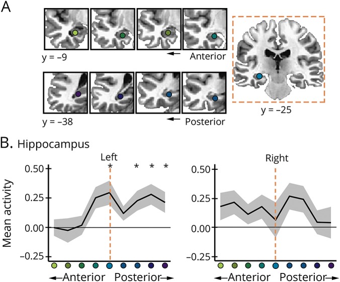
Figure 3. Stimulation increased hippocampal activity during memory formation.

(A) Segments along the anterior-posterior long axes of the left and right hippocampus relative to the average a priori target in the left hippocampus (y = −25). (B) Mean difference in recollection fMRI activity for each segment post-stim minus post-sham. Error bars indicate SEM. *p ≤ 0.05.
