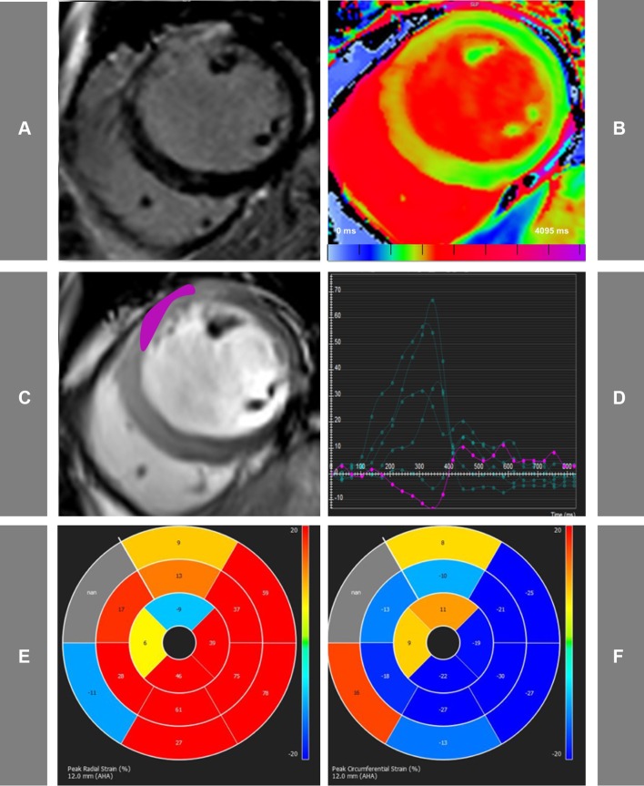
Fig. 1.
Representative acute CMR images used for the assessment of STEMI patients. Matching mid ventricular short axis slices acquired using LGE PSIR images (a), native T1-mapping (b), functional cine imaging (c). d The cvi42-derived tissue tracking analysis, whilst e and f display representative bull’s eye maps for radial and circumferential strain assessment, respectively. In this case of acute anterior myocardial infarction, the myocardium co-localized with the enhanced areas on LGE images, show prolonged T1 values and abnormal peak radial (Err) and circumferential (Ecc) strain

