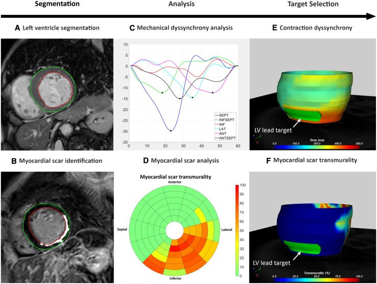
Fig. 3.
CARTBox workflow in images. a Segmentation of left ventricle. b Myocardial scar detected on CMR LGE scans. c Contraction timing analysis displaying delayed contraction of anterior and lateral segments. d Transmurality of scar showing inferolateral infarct of the left ventricle. e, f 3D-model of contraction timing (e) and scar transmurality (f) with manual selected target segment (green). ANT anterior, ANTSEPT anteroseptal, INF inferior, INFSEPT inferoseptal, LAT lateral, LV left ventricle, SEPT septal

