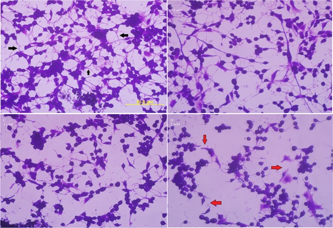
Figure 3.
Effect of chronic METH on cell morphology. C6 astroglia-like cells were treated with equal volume of vehicle (PBS control, A) or 0.5 (B) or 1 (C) or 2 mM METH (D) for 48 h. Morphological images of crystal violet dye stained cells were taken using an inverted phase contrast IX-70 Olympus microscope with a 40x objective lens. Inter-cellular connections in control cells (A) are shown by black arrows, while their loss in 2 mM treated cells (D) is shown with red arrows. Scale bar: 20 μm.

