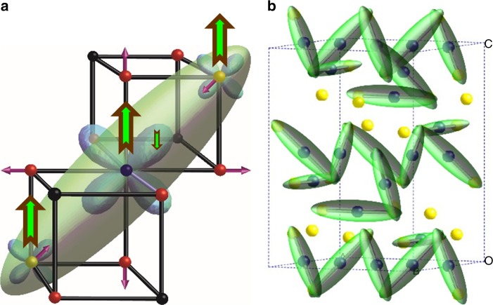
Fig. 4.
Trimeron bonding driven by magnetic order in magnetite. Charge-ordered Fe2+/Fe3+ states are shown as blue/yellow spheres, trimerons are green, and oxide ions are red. a A single trimeron unit consisting of three Fe sites with parallel S = 5/2 spins as shown by the brown–green arrows. Orbital order at the central Fe2+ site localises an antiparallel spin electron in one of the t2g orbitals, which distorts the local structure through elongation of four Fe–O bonds and shortening of the distances through weak bonding to two Fe neighbours in the same plane, as indicated by the purple arrows. The minority spin electron density is approximated by the ellipsoid shown. b Long-range order of trimerons in the monoclinic superstructure formed below the Verwey transition. Corner-sharing of trimerons results in a complex pattern of atomic displacements that has been used to model the local structure in the pair distribution functions

