Fig. 3.
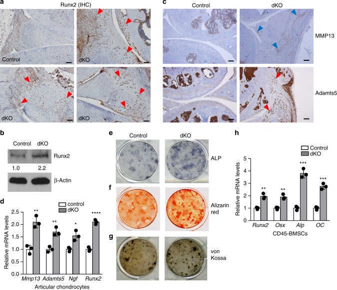
Deficiency of miR-204/-211 upregulates Runx2, cartilage-catabolic enzymes and osteogenic markers. a IHC results show enhanced Runx2 expression in the synovium, articular cartilage (AC), and meniscus of knee joints of dKO mice. Red arrowheads, Runx2-positive cells. Scale bar, 50 μm. b Western blot analysis shows Runx2 accumulation in CD45− BMSCs isolated from dKO mice. The numbers below the blot indicate the relative protein levels of Runx2 (normalized to β-actin) in each group compared to the control. n = 3. c IHC results of MMP13 and Adamts5 in control and dKO mice. Arrowheads, IHC-positive cells. Scale bar, 100 μm. d qRT-PCR analysis of Mmp13, Adamts5, nerve growth factor (NGF), and Runx2 expression in articular chondrocytes from control or dKO mice. Unpaired Student’s t test. n = 3. Data are shown as the mean ± s.d. e−g Alkaline phosphatase (ALP) staining (e), Alizarin red staining (f), and von Kossa staining (g) of BMSCs isolated from control or dKO mice. n = 3. h qRT-PCR analysis of Runx2, Osterix (Osx), Alp and Osteocalcin (OC) expression in CD45− BMSCs from control or dKO mice. Unpaired Student’s t test. n = 3. Data are shown as the mean ± s.d. *P < 0.05, **P < 0.01, ***P < 0.001, ****P < 0.0001. Uncropped western blot scans are shown in Supplementary Data 1

