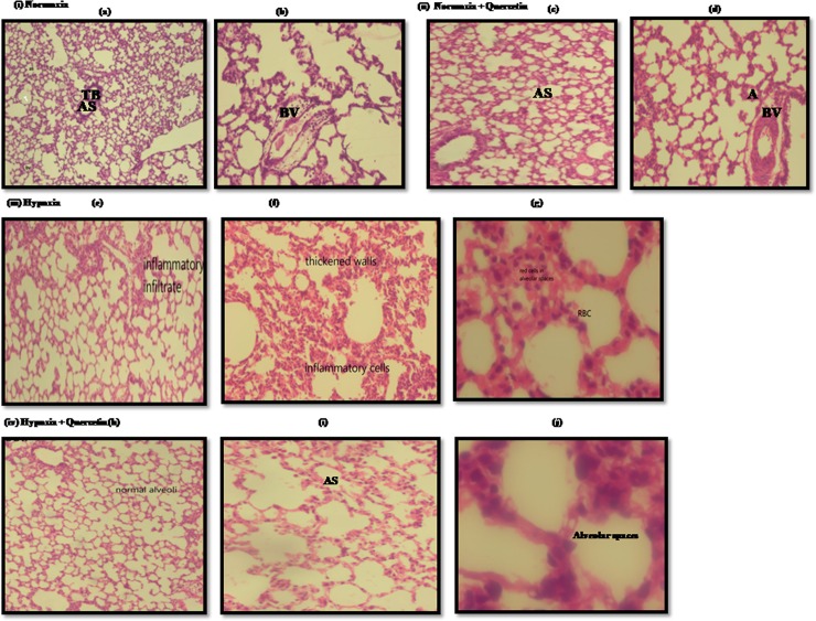
Fig 10. Histopathological images representing the collapsed alveoli, infiltration of inflammatory cells and RBCs, ruptured blood vessels and thin septae in lung of rats exposed to hypoxia at 7,620m for 6h.
(i) (a) and (b) Low power photomicrograph (10X) of lung section from normoxia control group (0h) representing normal lung parenchyma, clear AS and normal TB(ii) (c) Low power photomicrography of lung parenchyma of normoxic animal fed with Quercetin (50mg/kg BW), showed normal lung configuration with medium sized alveoli and normal blood vessels (d) High power photomicrography (40X) from the same section, exhibited normal alveolar spaces surrounded by medium septae (iii) (e) Low power photomicrograph of (10X) of lung section of hypoxic animal (hypoxia control (6h)) demonstrated the infiltration of infiltatory cells (f) High power photomicrographs (40X) of another lung section of hypoxic animal exhibiting the inflammation and thickened walls (g) High power photomicrograph (100X) of another lung section demonstrated the appearance of RBCs in alveolar spaces (iv) (h) Low power photomicrograph (10X) of lung parenchyma of hypoxic animal supplemented with quercetin (50mg/kg BW) showed normal alveolar structure (i) High power photomicrograph (40X) from the same section showing clear alveolar spaces and blood vessels (j) High power photomicrograph (100X) from the same section of hypoxia +Quercetin animal lung, manifested clear alveolar spaces devoid of any appearance of inflammatory cells and RBCs.where A- alveoli, AS- alveolar spaces, TB- terminal bronchiole, ICs- Inflammatory cells.

