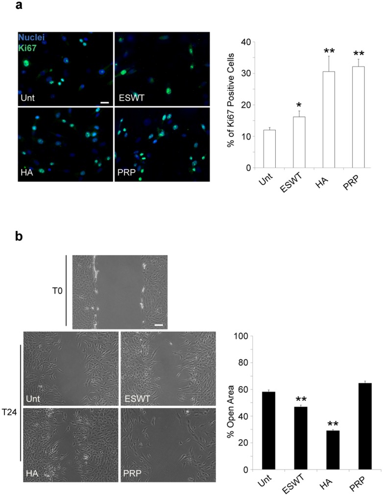
Fig 5. Proliferation and repair is stimulated by ESWT, HA and PRP treatment in chondrocyte cultures.

Analysis of proliferation and migration induced by ESWT, HA and PRP were performed as described in material and methods. (a) ESWT, HA and PRP effects on cell proliferation of chondrocyte cultures. Immunolabeling with anti-Ki67 antibody (green) was achieved on cell cultures after ESW, HA and PRP exposure for 24 days (T24). Nuclei are stained with DAPI. Photomicrographs are representative of one single culture. Bar 20 μm.Quantitative immunofluorescence analysis of proliferation induced by ESWT, HA and PRP was performed as described in material and methods. Results are shown as means ±SD. Student’s t-test was performed, and significance levels have been defined as p<0.001 (**) and p<0.05 (*). (b) ESWT, HA and PRP effects on cell migration of chondrocyte cultures. Results reported in the graph were obtained from four different cultures and represent the mean percentage ±SD of a residual open area as described in material and methods. Results are shown as means percentage ±SD. Student’s t-test was performed, and significance levels have been defined as p<0.001 (**) and p<0.05 (*). Bar 50 μm.
