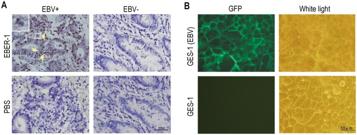
Fig. 1.
The detection of EBV infection in GC tissues and the GES-1 cell. (A) EBV genome detection in GC specimens by EBER-1 in situ hybridization (ISH) (magnification, 400 ×). Two cases of tissues showed to be EBV-positive (EBV +) and EBV-negative (EBV-) respectively. The cell-in-cell structures are indicated by yellow arrows. A magnified image is showed at the upper left corner. (B) The GFP expression in GES-1 cells post-infection of “cell-in-cell”. The fluorescence was observed at 48 h post-infection under a fluorescence microscope.

