Fig. 2.
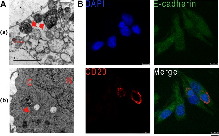
EBV observation and detection in gastric epithelial cell co-culture with EBV positive Akata cell. (A) The observation of Akata-EBV infection in GES-1 cells by electronic microscopy. (a) EBV-bearing Akata cells penetrated into GES-1. (b) Viruses were released from Akata into the cytoplasm of GES-1. N represents the nucleus, C represents the cytoplasm and red arrows indicate EBV-containing Akata cells. (B) Detection of EBV-positive Akata cells in GES-1 cells by using immunofluorecence assay. CD20 antibody was used for the detection indicating the membrane of Akata cells (red). E-cadherin staining (green) indicates the cell outline, and DAPI staining (blue) indicates the nucleus. A confocal microscope was used for the observation and image-taking. Scale bar, 10 μm.

