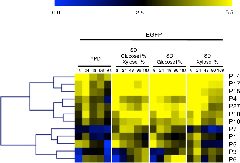
Fig. 2.
Expression of EGFP from monodirectional promoters. Fluorescence was measured by flow cytometry of cell populations sampled at five time points from four different media for strains harboring promoter constructs in orientation 1. The heatmap was generated using MultiExperiment Viewer (MeV), and promoters were clustered by hierarchical clustering using Euclidean distance. Fluorescence expression values are on a Log2 scale (color scale is shown in the upper bar), calculated as described in “Methods”. Each column represents a time point, shown in hours. Each row represents a promoter, named on the right side of the heatmap

