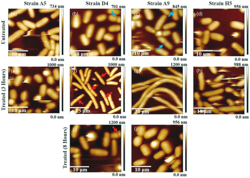
Fig. 2.
(a–j) Three-dimensional AFM height images. (a–d) Untreated MDR-E. coli cells (control), and (e–h) and (i, j) MDR-E. coli cells exposed to ampicillin at 3 and 8 h, respectively. The red arrowheads indicate elongated and dividing cells in presence of antibiotics and the blue arrowheads depict dividing cells in the absence of ampicillin.

