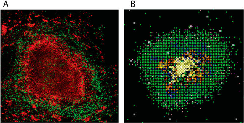
Figure 1.
Non-human primate an. simulated granulomas. Panel (A) Non-human primate granuloma stained for macrophages (Red CD68) and T cells (Green CD3). The very center (black, no staining) is caseous necrosis (Image courtesy of Dr. Josh Mattila, University of Pittsburgh). Panel (B) GranSim outputs. 2D snapshot, zoomed in to show detail showing macrophages (green-resting, blue-activated, orange-infected, red-chronically infected), T cells (pink IFN-g producing, purple-cytotoxic, white-Tregs), extracellular bacteria (yellow), necrosis (brown areas).

