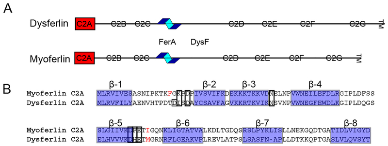
Figure 1:
A. Domain schematic of the dysferlin and myoferlin proteins. C2 domains are shown as boxes, FerA is depicted as a helical cartoon. The domains highlighted in red are the two C2A domains under consideration B. Structure-based alignment of dysferlin C2A and myoferlin C2A [31].β-strands are labeled above and highlighted as blue-shaded boxes. Residues observed to coordinate Ca2+ are boxed. Hydrophobic residues at the Ca2+ binding site that interact with the membrane are shown in red.

