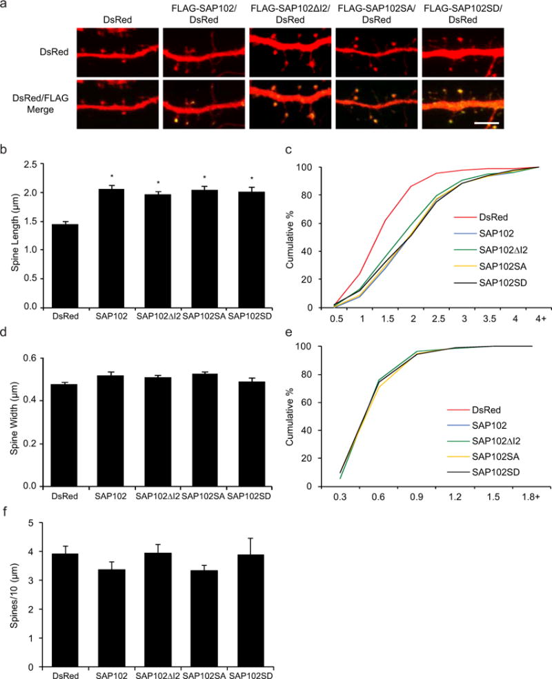
Figure 3. Phosphorylation of SAP102 on Ser632 doesn’t affect spine morphology.

a, Primary hippocampal neurons (DIV12) were transfected with FLAG-SAP102/DsRed, FLAG-SAP102ΔI2/DsRed, FLAG-SAP102 S632A/DsRed, FLAG-SAP102 S632D/DsRed or DsRed only. At DIV14, neurons were fixed and labeled with anti-FLAG antibody (green). Scale bar, 5 μm. b and d, Dendritic spine length and width were quantified by measuring DsRed signal using ImageJ software. c and e, Cumulative frequency plots of spine length and spine width are shown. f, Quantification of spine density (number of spines per 10 μm of dendrite length) in neurons as defined by DsRed expression. Data represent means ± S.E. (n = 10 neurons per condition from 3 independent cultures; 20 – 30 spines/neuron; *, p < 0.05, t test with Bonferroni’s correction after ANOVA).
