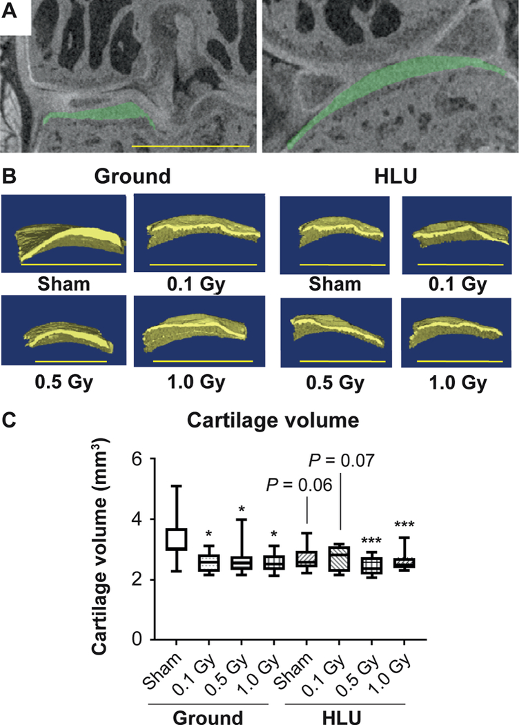
FIG. 1.

The volume of cartilage lining the medial tibial plateau is reduced compared to ground-sham after HLU and/or low-dose irradiation. Panel A: Region of interest indicating articular cartilage lining the medial tibial plateau (green). Panel B: Representative reconstructed 3D images of cartilage lining the tibial plateau used for biometric analysis. Panel C: Cartilage volume measurements in treatment groups (n = 10/group) compared using two-way ANOVA, with P values from Tukey’s post hoc analysis shown (*P < 0.05, **P < 0.01, ***P < 0.001). Scale bar = 500 μm. Other than treatments vs. ground-sham, no inter-group comparisons showed significant differences.
