Fig. 2.
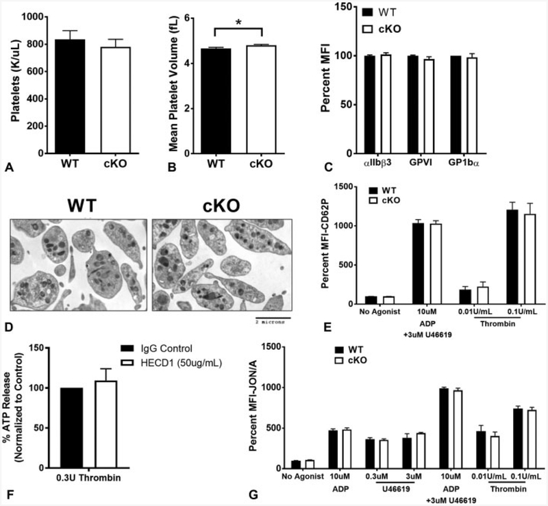
Thrombocytopoiesis and granule secretion are not affected by epithelial (E)-cadherin. (A) Platelet counts in wild-type (WT) (n = 12) and conditional knockout (cKO) (n = 20) animals. Average absolute values ± standard deviation (SD) are shown. (B) Mean platelet volume between WT (n = 12) and cKO (n = 20) animals. There is a small, but significant increase in mean platelet volume in cKO mice (p < 0.05). Average absolute values ± SD are represented. (C) Cell surface expression by flow cytometry of platelet-critical in tegrins and glycoproteins. Data are presented as per cent mean fluorescence intensity (MFI) normalized to WT ± SD(n ≥ 6). (D) Electron microscopy images of resting platelets from WT and cKO mice show no obvious differences in granule number or distribution. (E) Alpha granule release in response to indicated agonists was measured by cell surface expression of P-selectin (CD62P). Data are presented as percent MFI normalized to WT without agonist ± SD (n ≥ 7). (F) Dense granule release in human platelets inhibited with an antibody against E-cadherin (HECD1) in response to thrombin was measured by cell adenosine triphosphate (ATP) release. Data are presented as per cent ATP release normalized to control-treated platelets ± SD(n = 3). (G) Washed platelets were stim ulated with the indicated concentrations of adenosine diphosphate (ADP), U46619 or thrombin and activation assessed via activated αIIbβ3 (JON/A). Data are presented as per cent MFI normalized to WT without agonist ± SD (n ≥ 6).

