Figure 1.
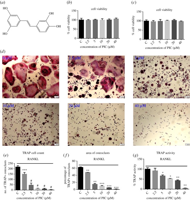
PIC attenuates RANKL-induced osteoclast differentiation. (a) The chemical structure of PIC; RAW264.7 cells were treated with various concentrations of PIC (2.5, 5, 10, 20, 40 µM) or vehicle (0.1% DMSO) at the different time interval for 48 h (b) and 72 h (c), and CCK-8 was used to measure cell viability; (d) RAW264.7 cells were differentiated into osteoclasts in the presence of M-CSF (10 ng ml−1), RANKL (20 ng ml−1) and PIC (0, 2.5, 5, 10, 20, 40 µM) for 4 days. Then cells were fixed with 4% paraformaldehyde for 20 min and stained by TRAP; (e) TRAP-positive cells in a well; (f) the area of TRAP-positive multinucleated cells; (g) TRAP activity quantification (*p < 0.05, **p < 0.01, ***p < 0.001, #p < 0.0001).

