Figure 3.

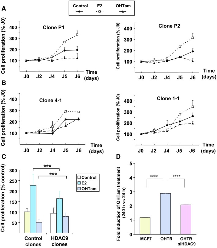
HDAC9 and the regulation of breast cancer cell proliferation by ER ligands. (A) Control MCF7 cell clones (empty vector; P1 and P2) were cultured in DCC medium supplemented with E2, OHTam or solvent alone (Control) for 5 days. Cell proliferation was measured with the MTT assay at days 2, 4, 5 and 6 and expressed relative to the absorbance at day 0. Results are the mean ± SD of three independent experiments, performed in triplicates. (B) The same as in A, but with MCF7‐HDAC9 cell clones (4‐1 and 1‐1). (C) Data obtained for each condition (control and MCFT‐HDAC9 cell clones) were pooled and compared. Data represent the mean ± SD of three independent experiments for each clone; ***P < 0.001 (Mann–Whitney test). (D) The growth of MCF7 and MCF7‐OHTR cells (transfected or not with a siRNA directed against HDAC9) was monitored using the XCELLigence system for a total duration of 10 days. The cell index corresponding to the number of viable cells was determined. The results represent, for each condition, cell proliferation measured after 240 h of culture in the presence of OHTam normalized by the values measured at 24 h (means ± SD; two independent experiments). ****P < 0.0001 (Mann‐Whitney test).
