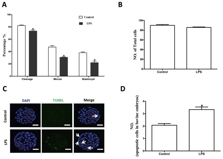
Figure 6.
LPS exposure decreases the parthenogenetic development of bovine oocytes. (A) Rates of different stages of parthenogenetic development after oocytes were preincubated with LPS for 22 h during in vitro maturation. (B) Total number of cells in blastocysts from each treatment group. (C) Representative photomicrographs. Blue, chromatin; Cyan, the transferase-mediated dUTP nick end labeling (TUNEL)-positive apoptotic nuclei. Bar = 20 μm. LPS, lipopolysaccharide. (D) Apoptotic cell rate. Asterisk indicates significant difference (p<0.05).

