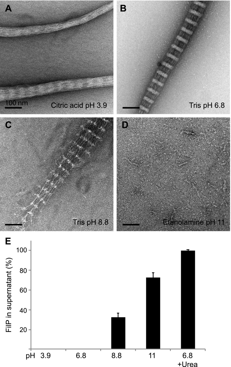
Figure 4. FilP filament formation is affected by pH.
(A) Negative staining EM images of FilP dialyzed in citric acid, pH 3.9, resulted in thicker filaments with a diffused banding pattern (e.g., the lower filament in this image) and condensed thinner filaments without striations (e.g., the upper filament in this image). (B) FilP dialyzed in standard Tris assembly buffer, pH 6.8, formed paracrystals with a banding pattern. (C) FilP dialyzed in Tris, pH 8.8, formed less compact striated filaments. (D) FilP dialyzed in ethanolamine, pH 11, formed rod-like structures. (E) FilP solubility measured by ultracentrifugation after FilP dialysis in buffers with pH ranging from 3.9 to 11. Bars represent protein content in the supernatant. The average of three experiments is shown, and error bars represent the SD, P ≤ 0.05, which was calculated using a one-way ANOVA test.

