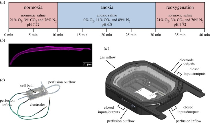
Figure 1.
Experimental protocol and equipment for subjecting turtle cardiomyocytes to an anoxic challenge. (a) Ventricular cardiomyocytes from juvenile turtles that embryonically developed in normoxia (21% O2) or chronic hypoxia (10% O2) were subjected to a 40-min experimental protocol to determine the effects of simulated normoxia, anoxia and reoxygenation (same treatment as normoxia) on cell shortening, [Ca2+]i, pHi and ROS production. (b) Isolated cardiomyocytes (image collected from a confocal microscope) were placed onto (c) a closed-cell bath, within (d) a custom-built chamber, in which the cardiomyocytes were subjected to the two treatments. Images courtesy of Warner Instruments (model RC-21BRFS) and Okolab (custom-built bold-line top stage incubator). (Online version in colour.)

